Lead Clinician, Vista Smile Studio
Published 1 June 2026
10 min read
Turkey teeth filed down to peg-like stumps describe one specific outcome of aggressive veneer or crown preparation, not a uniform clinical standard. Conservative porcelain veneer preparation removes 0.3–0.7 mm of enamel from the facial surface, preserving the bonded substrate that gives the restoration its 10–15 year survival profile. Vista Smile Studio applies a biomimetic protocol with a silicone reduction guide and digital wax-up, targeting maximum 0.7 mm facial reduction. The figures, the risks, and the UK regulatory framework that governs consent are covered below.
What “filed down” actually means in veneer preparation
The phrase "filed down" covers two clinically distinct procedures with very different biological cost. Conservative veneer preparation removes a thin band of enamel from the facial surface and a controlled wrap into the proximal contacts; full-coverage crown preparation removes the entire enamel envelope and exposes dentin around the whole circumference of the tooth.
Facial reduction vs full-coverage reduction
For a porcelain laminate veneer, the standard reduction is confined to the labial surface and the incisal edge. Facial reduction targets 0.3–0.7 mm depending on ceramic system, with a small chamfer at the cervical margin and an incisal reduction of 1.0–1.5 mm where shortening is planned.1 Lingual surfaces are usually left intact. By contrast, full-coverage crown preparation reduces axially around the entire tooth, removing 1.0–2.0 mm of structure and exposing dentin on every face. The visible "peg" silhouette in some Turkey teeth photographs is a crown preparation, not a veneer preparation.
Enamel preservation and bond strength
The clinical reason to preserve enamel is bond strength. Resin-cement bond to etched enamel reaches ≥20 MPa with contemporary adhesive systems; bond to freshly cut dentin reaches 10–15 MPa under the same conditions and degrades faster over time.2 A veneer cemented predominantly to enamel resists shear and fatigue load substantially better than one cemented predominantly to dentin. Where preparation strays into dentin across most of the bonded surface, the survival profile changes from a veneer's curve to something closer to a cemented crown.
Prep depth by material: a quantified table
Reduction depths track the ceramic system in use. Feldspathic porcelain is the most translucent and the thinnest workable layer; lithium disilicate trades a small amount of translucency for substantially higher flexural strength; zirconia is reserved for full-coverage crowns where load demands or aesthetic compromise allow.
Feldspathic porcelain (0.3–0.5 mm)
Feldspathic stacked-ceramic veneers permit the most conservative facial reduction at 0.3–0.5 mm. The thinness restricts the system's ability to mask underlying discolouration; the indication is typically minor reshaping or shade refinement on already light-shade teeth.
Lithium disilicate (0.5–0.7 mm)
Lithium disilicate (e.g. IPS e.max) is the contemporary workhorse for porcelain laminate veneers. Facial reduction sits at 0.5–0.7 mm with 1.0–1.5 mm incisal reduction. Flexural strength of approximately 360–400 MPa allows thinner restorations than zirconia while still covering moderate underlying discolouration.1
Zirconia full-coverage crowns (1.0–2.0 mm)
Zirconia is a crown material rather than a veneer material in routine cosmetic indications. Axial reduction of 1.0–1.5 mm and occlusal reduction of 1.5–2.0 mm are typical. The biological cost is loss of the entire enamel envelope; the indication is structural — heavily restored teeth, severe discolouration where masking demands opacity, or full-arch rehabilitation.
| Facial reduction | Incisal reduction | Axial reduction | Finish line | |
|---|---|---|---|---|
| Feldspathic porcelain | 0.3–0.5 mm | 1.0 mm | Not required | Light chamfer, equigingival |
| Lithium disilicate | 0.5–0.7 mm | 1.0–1.5 mm | Not required | Chamfer, equi- to slightly subgingival |
| Zirconia crown | 1.0–1.5 mm | 1.5–2.0 mm | 1.0–1.5 mm circumferential | Heavy chamfer or shoulder |
The Magne biomimetic preparation protocol
Pascal Magne's biomimetic framework formalised the case for enamel preservation and immediate dentin sealing in adhesive restorative dentistry. Its principles dominate the modern minimally invasive veneer literature.3
Silicone reduction guide and digital wax-up
Preparation begins with a diagnostic wax-up — physical or digital — that reproduces the planned final shape. A silicone matrix is taken over the wax-up and sectioned. The matrix is reseated on the teeth during preparation and used to verify reduction depth at every stage. Without a guide, the operator relies on visual estimation; with a guide, depth is mechanically referenced and over-reduction is detected before it occurs.
Immediate dentin sealing
Where preparation locally exposes dentin, the Magne protocol applies a dentin-bonding agent immediately, before impression. The freshly cut dentin is sealed against bacterial ingress, the hybrid layer matures over the temporary phase, and the final cementation bond performs more predictably. Delaying dentin sealing to the cementation appointment gives a measurably weaker bond and increases post-operative sensitivity.3
What happens if a tooth is shaved down too much
Pulpitis incidence and prep depth
Reported irreversible pulpitis after restorative preparation rises with depth. Conservative veneer preparation that stays within enamel reports incidence around 1%. Preparation that reaches dentin and approaches the pulp — including the circumferential reduction characteristic of crown preparation — reports irreversible pulpitis between 11% and 22% across the published clinical literature.4 The mechanism is bacterial ingress through cut dentinal tubules combined with thermal and mechanical insult to the pulp; the deeper the cut, the closer the tubules sit to vital tissue and the wider the diameter of each tubule.
When the tooth becomes a crown candidate, not a veneer candidate
Once axial reduction has reached dentin around the full circumference of the tooth, the substrate for a bonded veneer is no longer present. The realistic restorative options become a full-coverage crown, an endodontic-then-crown sequence if the pulp has been compromised, or — where the residual structure is inadequate — extraction with implant placement. The decision is not aesthetic; it is biomechanical and biological.
Figures aggregate small clinical series; absolute incidence varies with operator, ceramic system and follow-up window. See Trushkowsky and Burke 2012.
| Category | Value (%) | Notes |
|---|---|---|
| Minimal prep (enamel only) | 1 | Conservative veneer protocol |
| Moderate prep (partial dentin) | 5 | Typical aggressive veneer prep |
| Aggressive prep (crown) | 11–22 | Circumferential reduction |
Eight red flags that signal aggressive preparation
The following indicators precede most failed cases reported by UK-registered dentists treating returners. They are observable by the patient before the final restoration is bonded.
- Peg-shaped temporaries. Provisional restorations sit on stumps that taper sharply toward the gum line — a crown preparation profile, not a veneer preparation profile.
- Dentin exposed across most of the prepared surface. A conservative veneer prep leaves visible enamel at the cervical margin and along the proximal walls; widespread yellow-tinged dentin is a crown prep.
- No diagnostic wax-up presented before preparation. Without a wax-up there is no reference for either the operator or the patient to evaluate the planned outcome.
- No silicone reduction index used during preparation. Reduction is then estimated by eye, increasing the risk of over-reduction in the deeper segments of the tooth.
- No shade preview or trial smile. Final shade is committed to before the patient has seen it in their own face.
- No written consent referencing irreversibility. The British Association of Cosmetic Dentistry's guidance on minimally invasive cosmetic dentistry treats irreversibility consent as foundational.5
- Compressed one-visit treatment for full-arch cases. Preparation, impression and cementation in a single appointment removes the planning checks built into a staged protocol.
- No record of pre-treatment intraoral scan or impression. No baseline exists for the original tooth shape, contour or shade.
UK consent standards under GDC Standard 3.1
The General Dental Council's Standards for the Dental Team Standard 3.1 sets the threshold for valid consent: information must be provided in a way the patient can understand, alternatives must be discussed, and the consent must be voluntary and specific to the proposed treatment.6 Standard 3.1 is the framework UK-registered dentists are accountable to, but its substance describes a baseline that any cosmetic dental clinic working with UK patients can reasonably be expected to honour. Where treatment is delivered abroad, UK patients retain their right to make a fully informed decision; the responsibility for delivering the underlying information rests with the treating clinician.
Practical implications for veneer preparation include: a written treatment plan listing the planned material and approximate reduction; a written acknowledgement that enamel removal is irreversible; an explicit description of the failure modes covered in any guarantee; and the alternatives — including no treatment, composite bonding, and orthodontic correction — that have been considered and ruled in or out.
Vista Smile Studio's conservative preparation protocol
Vista Smile Studio's veneer protocol is built around enamel preservation, mechanically referenced reduction, and written consent. The clinic operates from Didim (Altinkum), Aydın province, under International Health Tourism Authorization issued by the Turkish Ministry of Health.
Digital wax-up first
Every veneer case begins with a digital wax-up generated from the pre-treatment intraoral scan. The wax-up is shared with the patient before any preparation is performed and a 3D-printed mock-up is placed in the mouth at the consultation visit so the planned shape can be evaluated in context.
Intraoral check before bonding
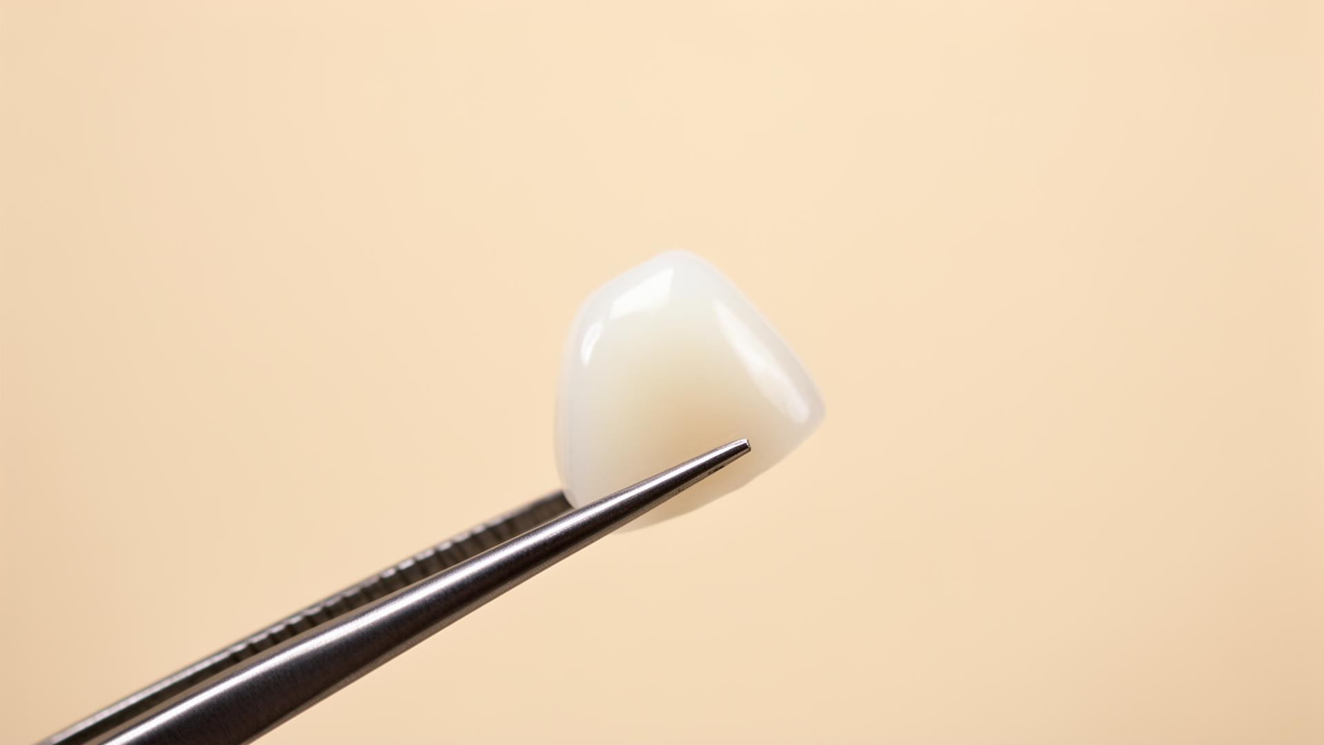
A silicone matrix made from the approved wax-up is sectioned and reseated over the prepared teeth at the end of the preparation phase. Reduction is verified at facial, incisal and proximal locations. The target facial reduction is ≤0.7 mm and an intact band of enamel is preserved at the cervical margin where the case allows. Vista's cosmetic work carries a 5-year guarantee covering defined failure modes, provided to the patient in writing at the time of treatment.
Restoring filed-down teeth: options when the damage is done
Where preparation has already over-reduced a tooth, the realistic restorative options narrow with the depth of the cut. Where enamel remains and the pulp is healthy, replacement with a new bonded veneer or partial-coverage ceramic onlay may be possible. Where the tooth has been circumferentially reduced into dentin, the appropriate restoration becomes a full-coverage crown. Where preparation has compromised the pulp, root canal treatment is performed before final restoration, and where residual tooth structure is inadequate to support any restoration, extraction and implant placement is considered. The decision tree is mapped out in detail in the companion article on Turkey teeth gone wrong, and Vista's veneer service page sets out the clinical assessment pathway.
Related Vista guides on the wider veneer journey: how long Turkey veneers last and what Turkey teeth are made of.
Frequently asked questions
Continue at Vista Smile Studio
Speak to our clinical team
References
- [1]Aschheim KW. Esthetic Dentistry: A Clinical Approach to Techniques and Materials. Elsevier, 4th edition. Reference text for prep-depth ranges by ceramic system.
- [2]Magne P, Belser UC. Bonded porcelain restorations: enamel preservation and adhesive performance. Quintessence Publishing.
- [3]Magne P. Immediate dentin sealing: a fundamental procedure for indirect bonded restorations. Journal of Esthetic and Restorative Dentistry, 2005. doi:10.1111/j.1708-8240.2005.tb00103.x
- [4]Trushkowsky RD. Pulpitis incidence following veneer preparation: a clinical review. Compendium of Continuing Education in Dentistry.
- [5]British Association of Cosmetic Dentistry (BACD). Position statement on minimally invasive cosmetic dentistry. https://bacd.com/
- [6]General Dental Council (UK). Standards for the Dental Team — Standard 3 (Obtain valid consent). https://www.gdc-uk.org/standards-guidance/standards-and-guidance/standards-for-the-dental-team
About the author
Dr. Yusuf Aydin, DDS
Lead Clinician, Vista Smile Studio
Dr. Yusuf Aydin is the lead clinician at Vista Smile Studio in Didim, Türkiye, with more than 15 years of experience in cosmetic and implant dentistry. He oversees treatment planning, surgical placement, and prosthetic delivery for international patients, with a particular focus on UK cases referred through Vista's two-trip treatment model.
View full author profile →